云端病例
Staged ROTA-ablation for the undilated LAD lesion during emergence PCI
Zhang Jun-Jie
Nanjing First Hospital
Nanjing Medical University
CASE SUMMARY
Patient Demographic
Age, 75 yrs.
Gender, Female
Clinical History
Repeated chest pain for 10-year, worsening for four days.
The details of hyperthyroidism are unavailable
Risk factors
Hyperlipidemia
Hypertention
Examination
Cr, 101.00 umol/L
TNI:2.36 ng/L
Diagnosis
NSTEMI
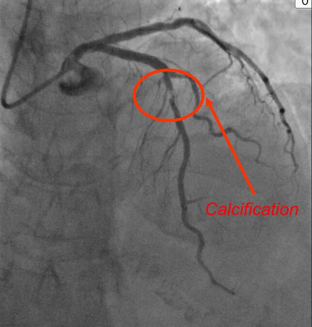
BASELINE ANGIOGRAPHY
(2016/06/21)
Failed to dilate mid-LAD lesion
stenting prox-LAD with 3.5/18 SES
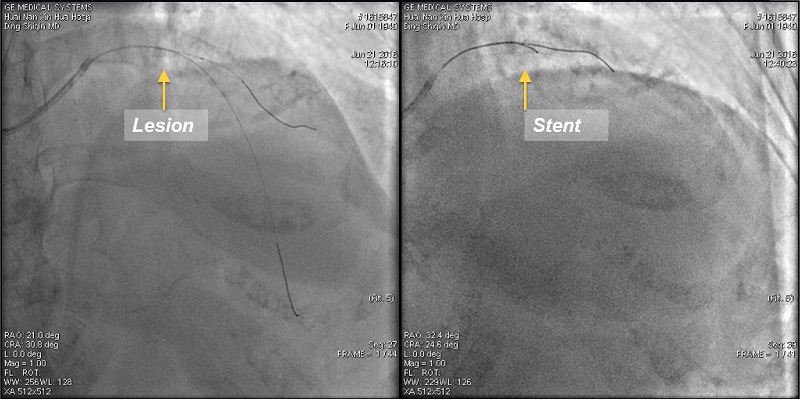
Lesion
Pre-dilation:1.5*15 Maverick, 16 atm
Stent
3.5/18 Firebird2 @12atm
Admittedto my hospital for UAP
(2016/7/7)
Implications of Coronary Calcification
Coronary calcification results in:
• Impaired stent delivery, decreased stent expansion, increased malapposition and edge dissections
• Decreased procedural success
• Increased procedural complications
• Increased rates of stent thrombosis and restenosis
StoneG. TCT 2016
What Tools do We Have to Detect Coronary Calcification
(in the cath lab)?
Imaging Coronary Calcification
• Fluoroscopy/cineangiography
• Intravascular ultrasound (gray-scale and radiofrequency)
• Optical coherence tomography
Pretreatment for LAD Calcification
GC: 6F EBU
GW: Runthough
Balloon: 1.5 * 12mm Sprinter
16 atm
2.0 * 8mm Quantum
16 atm can’t cross the lesion
Catheter: AtlantisTM SR Pro
Failed to place IVUS catheter in middle LAD
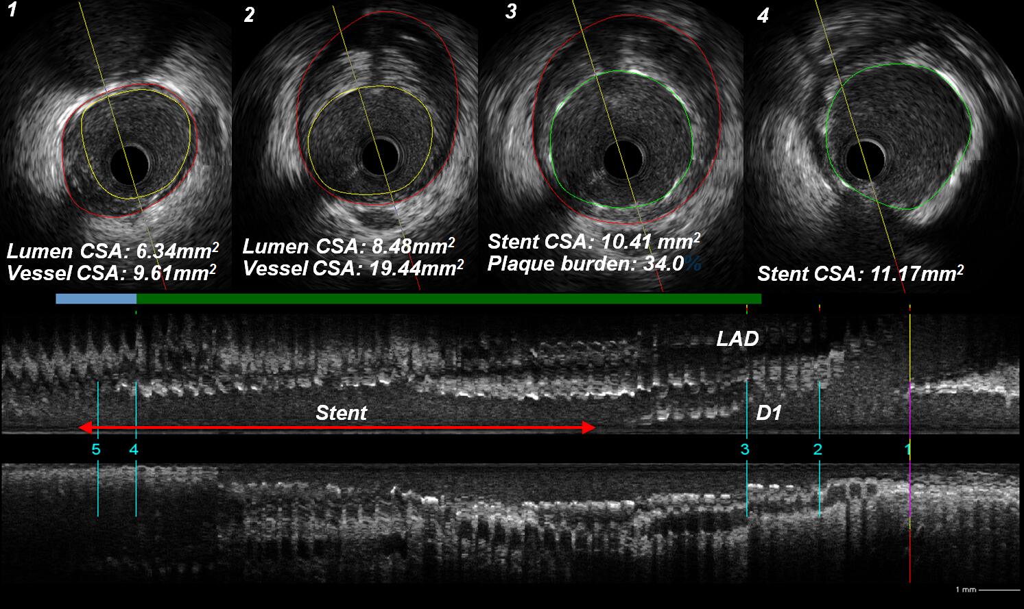
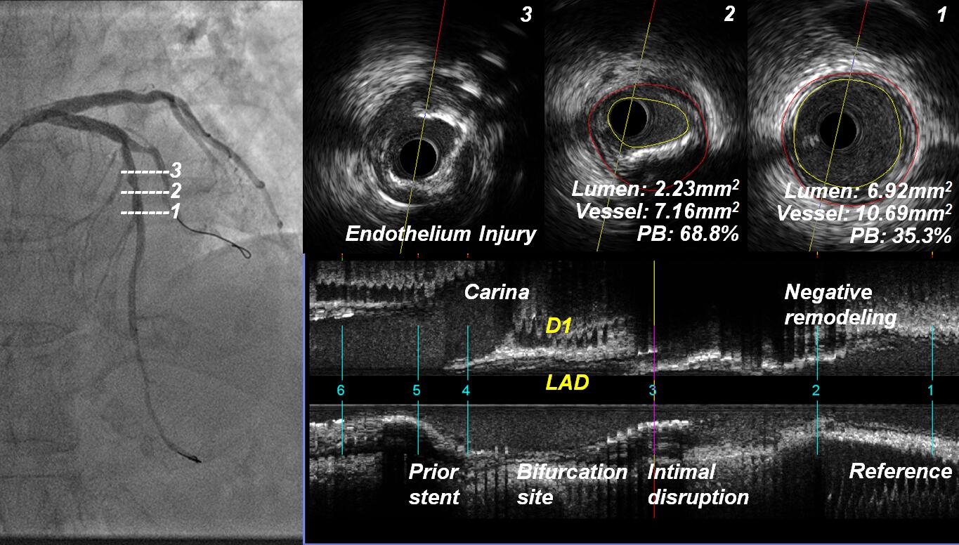
IVUS Finding
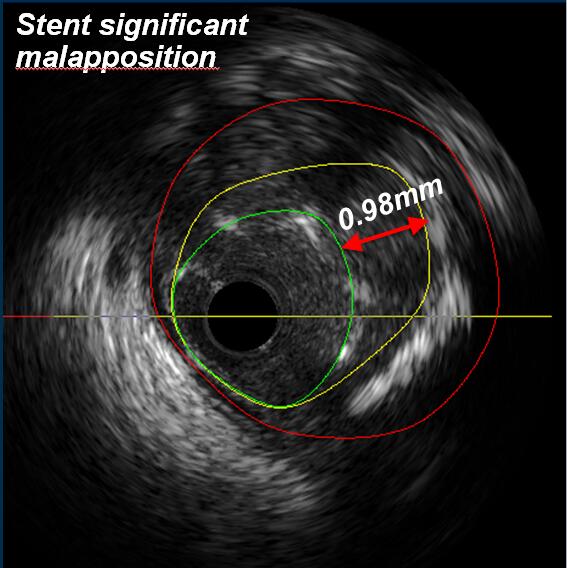
Lumen CSA: 9.80mm2
Stent CSA: 5.10mm2
Dilation with Non Compliance Balloon
3.5 * 8mm Quantum, 16 atm
IVUS Catheter: Atlantis™ SR Pro
IVUS Finding after Dilation
Pretreatment for LAD Calcification
WhatToolsdo We
Have to Treat
Coronary
Calcification
(inthe cath lab)?
Treatment of Calcified Lesions: Options
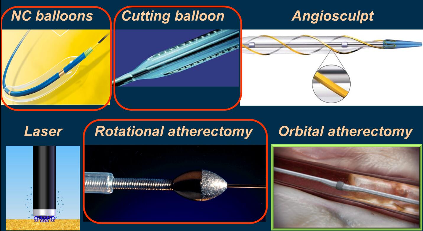
StoneG. TCT 2016
Treatment of Calcified Lesions:PCIguidelines
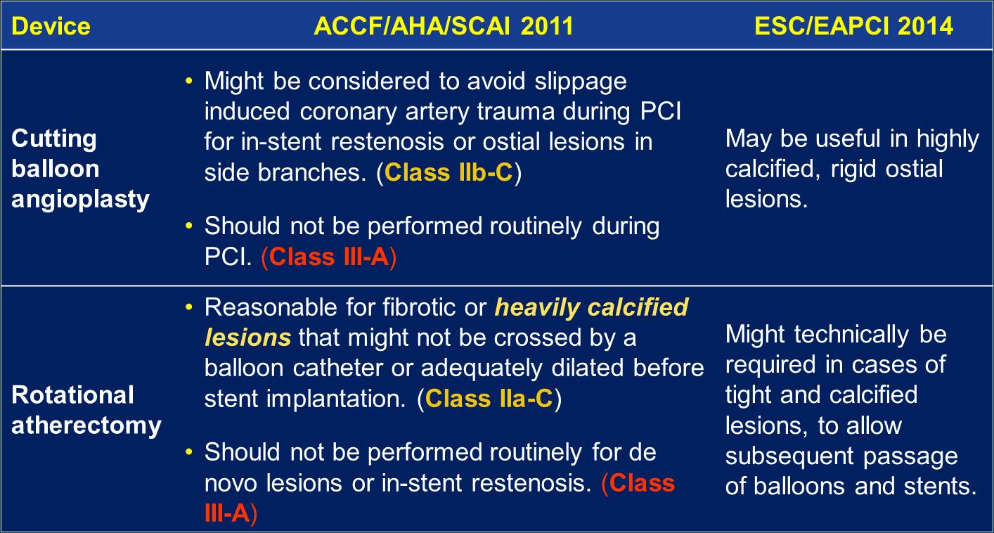
Levine GN et al. JACC 2011;58:e44-122
Windecker S et al. EHJ 2014;35:3541-619
Rotational Atherectomy
1.5mm burr,160,000rpm,20s@3r
Pre-ROTA
Post-ROTA
IVUS Finding after ROTA
Cutting Balloon Dilation following ROTA